The Scientist Behind It · Biology
The 100-Hour X-Ray Photograph That Revealed the Shape of DNA
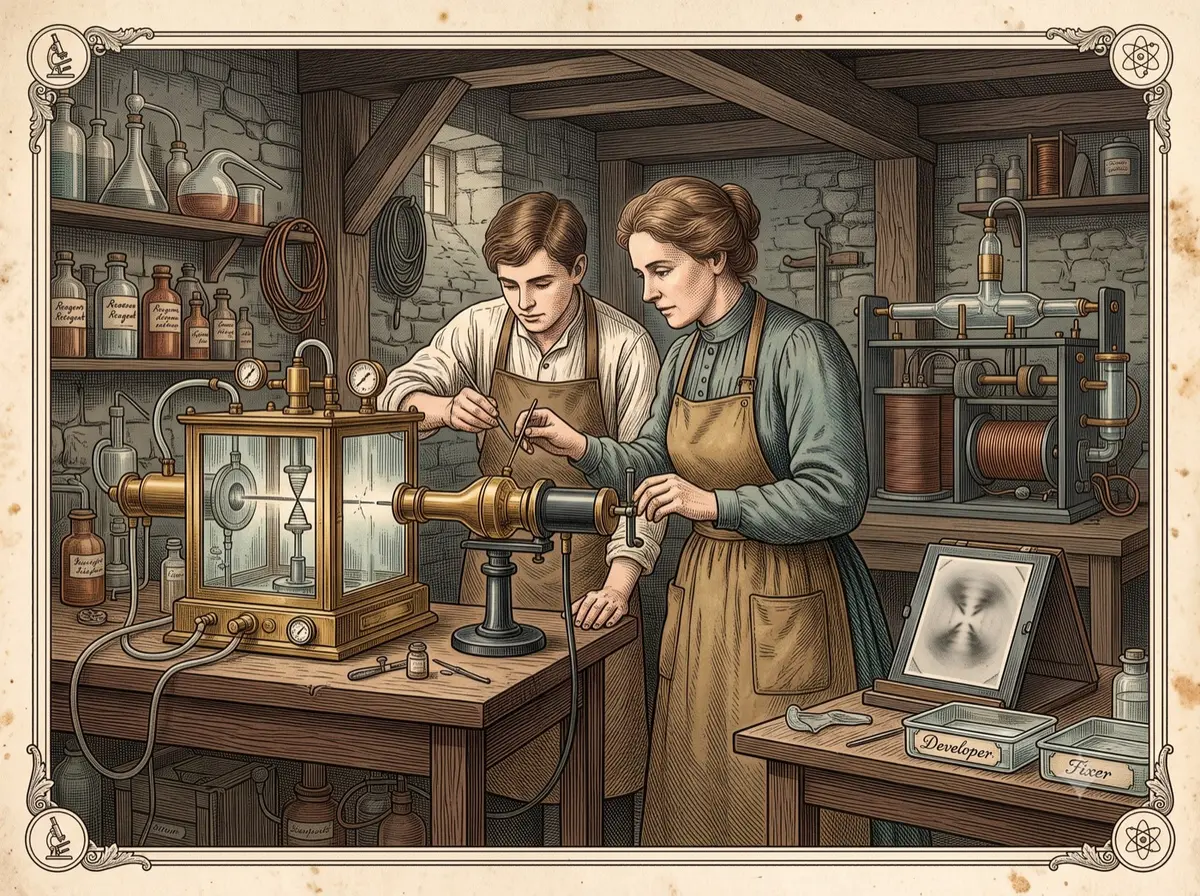
In a basement lab at King's College London in May 1952, a chemist named Rosalind Franklin and her graduate student Raymond Gosling clamped a DNA fiber no thicker than a strand of hair into a humidity-controlled chamber, aimed an X-ray beam at it, and walked away. For one hundred hours, invisible rays struck the fiber and scattered onto a photographic plate behind it. When the plate finally came out of the developer, it showed a dark, near-perfect X of smudged spots. Gosling logged it as the fifty-first diffraction photograph he'd taken. No one in that room could have predicted it would end up on a British coin.
By the Curiosities Desk · · 6 min read · Filed under Biology · Today’s round
Part of The Person Who Would Not Drop It
What happened: The X-Ray Photo of DNA
Rosalind Franklin was a physical chemist who had spent the late 1940s in Paris using X-ray crystallography — a technique that fires X-rays at a sample and reads the scattered pattern to infer the arrangement of atoms inside it — to study the structure of coal. In 1951 she moved to King’s College London to apply the same method to DNA, joining a biophysics unit led by John Randall, with Maurice Wilkins as deputy. Franklin and Wilkins never got along; their working relationship was tense from the start, in both temperament and approach.
Franklin made fast progress anyway. She discovered that a DNA fiber could exist in two distinct forms depending on humidity: a drier, crystalline “A” form that produced sharp, detailed diffraction patterns, and a wetter, more disordered “B” form that produced blurrier ones but was easier to interpret. Franklin devoted most of her energy to the harder, data-rich A form. She judged the B form a side issue, and by mid-1952, having failed to resolve the A form’s structure, she was doubting it was helical at all — she even wrote Wilkins a joke “death notice” mourning the demise of the DNA helix.
The B-form image that Gosling shot on May 2, 1952, at close to 92% relative humidity, later became known as Photograph 51. It showed a clean, symmetrical X — a classic signature of a helical structure. Franklin set it aside to keep working on the A form.
By early 1953, Franklin was preparing to leave King’s for Birkbeck College and had been told to leave her DNA work behind. Gosling, now under Wilkins’s supervision, passed him Photograph 51. On January 30, 1953, Cambridge researcher James Watson visited King’s, fresh off seeing a flawed DNA model that the chemist Linus Pauling had just circulated — a paper that had spurred Watson and his collaborator Francis Crick to resume their own model-building. After a tense exchange with Franklin, Watson was shown Photograph 51 by Wilkins, without asking Franklin first.
Around the same time, Crick’s supervisor, Max Perutz, passed Watson and Crick an internal report Franklin had written for a Medical Research Council committee visit. It contained her precise measurements: DNA’s repeating unit stacked every 34 angstroms (a unit of length equal to one ten-billionth of a meter), its crystal unit cell was unusually large, and its symmetry implied two strands running in opposite directions. Historians who examined this material later concluded that this data-dense report, not the single photograph, did more to steer Watson and Crick’s final model.
Working through roughly six weeks of model-building — described by both men as trial and error with cardboard and wire — Watson and Crick arrived at a double-helix structure. In April 1953, Nature published three papers back to back: Watson and Crick’s model, a supporting paper from Wilkins and colleagues, and one from Franklin and Gosling presenting their own X-ray evidence. Watson and Crick’s paper credited being “stimulated” by unpublished results from Wilkins and Franklin, though it understated how much of that data they had actually seen.
Why it was strange: The X-Ray Photo of DNA
A single photograph became mythologized as the moment DNA’s secret was cracked — commemorated on a British 50-pence coin, dramatized in a West End play starring Nicole Kidman, and repeated in classrooms as the instant Watson understood in a glance what Franklin supposedly couldn’t. But that version doesn’t hold up well. Even Watson and Crick agreed at the time that no single diffraction photograph could prove a structure on its own; other arrangements could in theory produce a similar pattern. Historians who dug into Franklin’s lab notebooks in 2023 found she understood far more about DNA’s helical nature than the popular story credits her with — she simply hadn’t yet made the final leaps linking her data to a complete double-helix model, in part because she was working largely alone, without the running dialogue Watson and Crick had with each other and with Wilkins.
The same thread — Genetics — continues in The Corn Geneticist Who Saw Genes Jump — And Waited 32 Years to Be Believed.
What scientists learned: The X-Ray Photo of DNA
Franklin’s measurements, combined with Watson and Crick’s modeling, revealed that DNA is a double helix: two strands of alternating sugar and phosphate twisted around each other like a twisted ladder, with the four chemical bases — adenine, thymine, cytosine, and guanine — paired inside, facing each other like rungs. The strands run in opposite directions, and the pairing rule (adenine with thymine, cytosine with guanine) means each strand carries enough information to reconstruct its partner. Franklin’s own notes from early 1953 record her independently reaching a version of the discovery’s deepest implication: because the sequence of bases along a strand didn’t affect the surrounding structure, “an infinite variety of nucleotide sequences would be possible” — meaning a single kind of molecule could encode the instructions for every living thing.
Elsewhere among living things: From Mendel’s Peas to CRISPR: A Short History of Genetics.
How it reaches us today: The X-Ray Photo of DNA
Every technology built on reading or editing genetic material — DNA sequencing, PCR testing, genetic ancestry kits, CRISPR gene editing, forensic DNA matching, prenatal genetic screening — depends on this structure holding true. Franklin’s own X-ray crystallography techniques also went on to reveal the structures of the tobacco mosaic virus and, at the time of her death, the polio virus, laying groundwork for structural virology still used today.
Another scientist, told from the human side: The Priest Who Put Tiny Taffeta Pants on Frogs to Find Out Where Babies Come From.
Fun fact: The X-Ray Photo of DNA
The “51” in Photograph 51 isn’t a code or a clue — it’s simply the fact that it was the fifty-first X-ray diffraction image Raymond Gosling had shot in the lab’s numbered sequence.
Sources: The X-Ray Photo of DNA
- Rosalind E. Franklin & Raymond G. Gosling, “Molecular Configuration in Sodium Thymonucleate,” Nature 171, 740–741 (1953) — https://doi.org/10.1038/171740a0
- James D. Watson & Francis H. C. Crick, “Molecular Structure of Nucleic Acids,” Nature 171, 737–738 (1953) — https://doi.org/10.1038/171737a0
- Matthew Cobb & Nathaniel Comfort, “What Rosalind Franklin Truly Contributed to the Discovery of DNA’s Structure,” Nature 616, 657–660 (2023) — https://www.nature.com/articles/d41586-023-01313-5
- “DNA X-ray Diffraction Image Known as Photo 51,” Science History Institute Digital Collections — https://digital.sciencehistory.org/works/skau3rn
- “How Rosalind Franklin’s ‘Photo 51’ Revealed DNA’s Structure,” HISTORY.com — https://www.history.com/articles/rosalind-franklin-photo-51-dna
This article mentions Rosalind Franklin’s death from ovarian cancer at age 37. If ovarian cancer has touched your life, organizations like the Ovarian Cancer Research Alliance offer information and support.
Related stories: The X-Ray Photo of DNA
- From Mendel’s Peas to CRISPR: A Short History of Genetics — Pea plants to gene editing in one long thread
- The Corn Geneticist Who Saw Genes Jump — And Waited 32 Years to Be Believed — She watched genes jump and waited 32 years for the field to agree
- The Graduate Student, the ‘Little Green Men,’ and the Nobel That Got Away — She found pulsars; the Nobel went to her supervisor